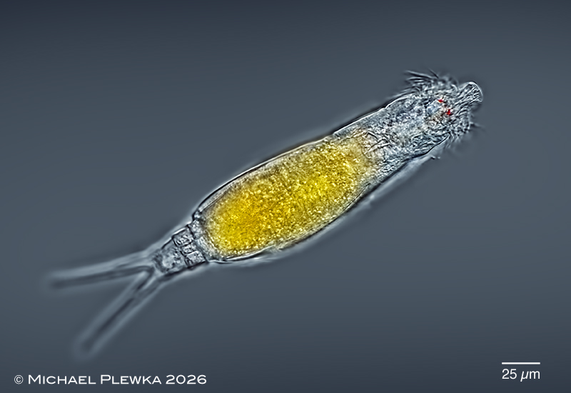
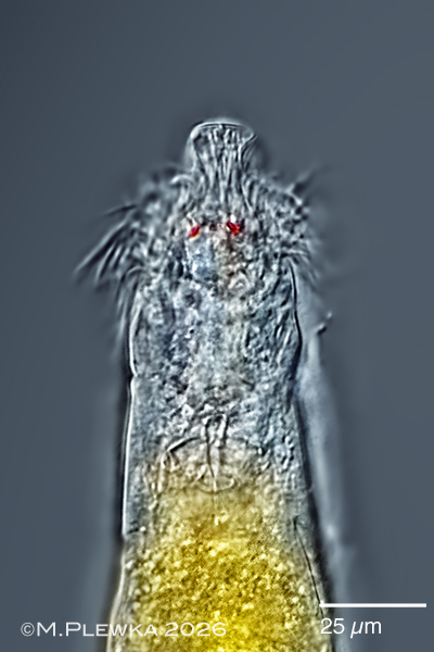
Dicranophorus hercules: dorsoventral view; focus plane on the lobed rostrum and the two red eyespots. The foot has 2 pseudosegments.
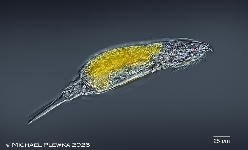
Dicranophorus hercules: lateral view. (1)
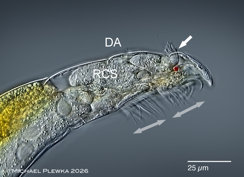
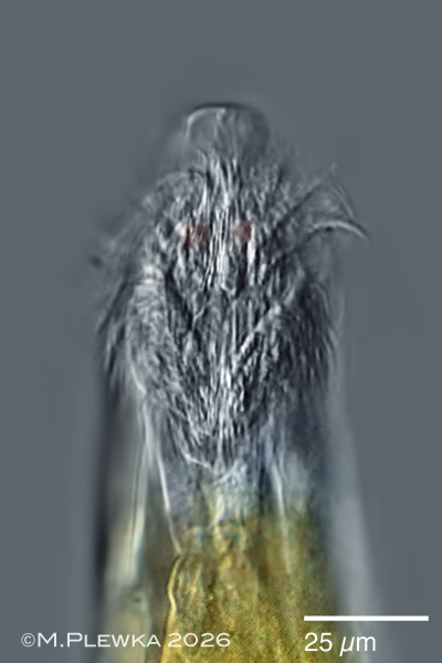
Dicranophorus hercules: lateral view of anterior part. Upper image: focal plane on the right red eyespot. The rotatory apparatus not only consists of the ventral ciliary field ( which seems to be divided in 2 parts (marked by double arrows), but also of lateral ciliary tufts (the right one marked by arrow) are visible. The unconspicuous dorsal antenna (DA) is faintly visible. The retrocerebral sac(RCS) is very large.
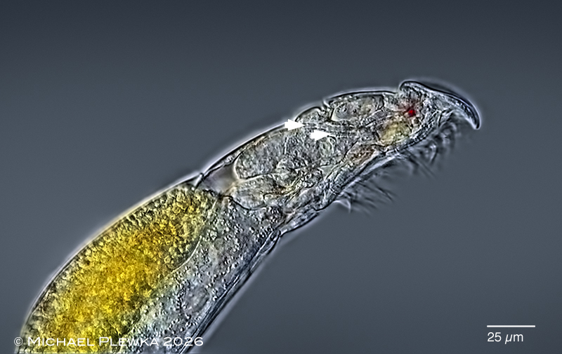
Lower image: the arrowheadss point to 2 structures that may be the ducts of the retrocerebral sac and the duct of the right subcerebral gland. Images not to scale. (2)
Dicranophorus hercules: two aspects of the anterior part. Left: dorsoventral view, optical transect; focal planeon the flexible rostrum which is laterally expanded at the tip and narrows head-wise. Red eyespots with light-refracting structures. Also part of the forcipate trophi are visible. Right: ventral view; focal plane on the ventral ciliary field. (1)
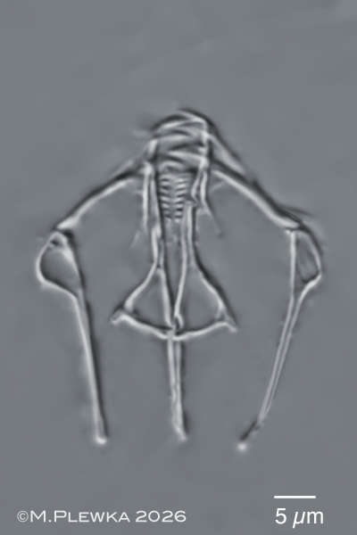
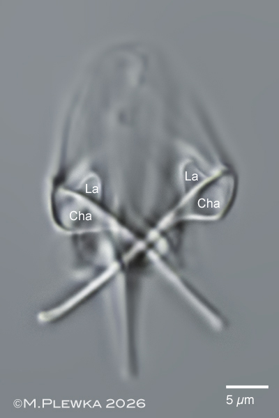
Dicranophorus hercules: left image: forcipate trophi; rami with spine-shaped alulae. The inner margins of the rami have ≈8 acute teeth of similar size. Right image: focus plane on the manubria with chamber (Cha) and lamella (La). Images not to scale. (1)